Grow&Mix Bioink
What
What if we could produce colors and plastic from bacteria in our kitchens? What if we could create materials, colors and machines for building our objects? DIY is the idea of doing by yourself those activities reserved to the industrial production system that encourages alienation and non-participation. Imagine people producing machines ink and biomaterials out of microorganisms found in Nature and brought to their kitchens. Project developed at Hackuarium with the collaboration of Prof. Xavier Perret. Conceptualized during my first semester at HEAD Geneva, the result was exhibited at Fiera del Mobile di Milano in 2015.
Who
Vanessa Lorenzo
Hackuarium team that helped to push this project forward: Sachiko Kana, Luc Henry, Yann Pierson, Gianpaolo Rando, Yann Heurtaux, Carmelo Bisognano (Univercité) and Clément Drévo.
Xavier Perret UNIGE that provided proof of concept experiment with the Violacein at his lab.
Naiyi Wang that designed infographics
Alexandra Midal and Mathieu Bassée HEAD Genève that promoted the early project in 2015.
How
Pr(ink)t plastic, it's fantastic!
Le challenge : construire un objet vivant, plein de bactéries que nous devons nourrir et soigner pour obtenir des couleurs riches pour les utiliser dans une imprimante 3D.
Symbiose et ailleurs. Serait-il très intéressant si est possible d'interagir dans ce système biomécanique avec tes propes bactéries. Expanded self sounds good to me ;)
Certaines bactéries naturelles et non pathogènes, comme le Janthinobacterium Lividum, font des pigments violet foncé. Un mélange de produits chimiques fournira différentes couleurs (c'est aussi possible de faire papier avec levure et acide acétique).
Il faudrait :
1.- Acheter les bactéries
2.- Modifier une imprimante 2D
3.- Système de transmission de bio-encre et un objet esthétique qui permettra de l'utiliser.
A la fin cela ressemblera a un petit laboratoire de bactéries sympathiques.
07/11/2014
Learn with Clément Drévo how to make a Control Petri-dish for tracking the growth of a contaminated one with my own finger bacteria.
12/11/2014
Learn with Gavrilo Bozovic the use of the UV and make tests with different exposure times 10' 30' 45' 60'
Check the results of the use of the UV in different petri dishes for different exposure times: not working properly, will use the autoclave when repaired.
26/11/2014
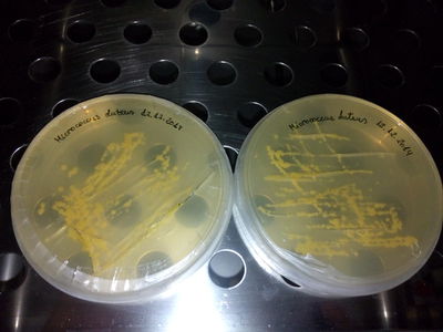
Select with Yann Pierson a bacteria which is actuallly producing Yellow color. And preparing together with Sachiko the LB medium and learn how to sterealize it in the"pressure cooker".
27/11/2014
All these strains produce pigments:
Micrococcus luteus (ML DSMZ) from DSMZ color: yellow Rhodococcus coprophilus (RC) from DSMZ color: pink Janthinobacterium lividum (JL) from DSMZ color: blue (interesting link http://schaechter.asmblog.org/schaechter/2009/04/by-jenna-tabor-godwin-rhona-stuart-rosa-i-león-zayas-and-chitra-rajakuberan--------janthinobacterium-cultured-on-nb-aga.html) Xanthomonas campestris (XC) from DSMZ color: yellow Rhizobium etli (RE) from DSMZ color: brown Vogesella indoferra (VI) from Dr. Simon Park color: dark blue Arthrobacter agilis (Aag) from Dr. Simon Park color: pink Chromobacterium violaceum (CV) from Dr. Simon Park color: purple Dermacoccus nishinomyaensis (DN) from Dr. Simon Park color: orange Micrococcus roseus (MR) from Dr. Simon Park color: salmon Arthobacter polychromogens (AG) from Dr. Simon Park color: dark blue Micrococcus luteus (ML) from Dr. Simon Park color: yellow
The purple pigment is produced by two of them: - Chromobacterium violaceum - Janthinobacterium lividum
Both produce Violacein. Do mind that C. violaceum is a level 2 organism, opportunistic pathogen. So. I guess the best option is the Janthinobacterium lividum.
Final strains, biosafety level 1: J. Lividum M. Luteus M. Roseus M. Aurantica
03/12/2014
--------------------------------------
Inspiring projects: http://www.ingridnijhoff.nl LaPaillase Grow your Ink The Waag Society
05/12/2014
LB medium (liquid, no agar) See LB protocol
Autoclave instructions: 0.-Plug 1.-fill the bottom with water till reach the 1st level/grid on the bottom. 2.-put your bottles on the top of the grid cubes (covered by another "plane" grid) 3.-close it but not to tight, close the little screw where it written "fermé". 4.-make sure you put 125°C and 1,5T. 5.-it takes 35 min to sterealize (20min to reach the heating t° and 15min to heat it up till sterealized) 6.-wait till is completely cooled down 7.-open (not open till it has reached the room t°) 8.-Unplug Finally extract the culture and empty a little quantity on the new erlenmeyer
- /
EXTRACTIONS: First take some culture from the original bottle, "source". Prepare the table: Clean it with alcohol Fire up the gas in order to have a clean atmosphere without air bacteria Remove the aluminium foil/protection from the mouth of the source bottle & heat it up a bit with fire the mouth of the bottle before emptying the "source" empty the source bottle on the new bottle heat it up a bit with fire the mouth of the bottle and the aluminium foil before putting in again the aluminium foil.
Then we continue the process with this tools:
1.- Pipette, always use the plastic cone(aeroject) on them: We extract some an put them in tubes (then close them).
2.- Then we shake the tubes in the automatic vortex at 10 x 1000 and 10 minutes
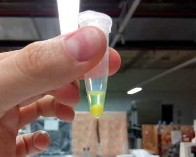
3.- Empty the lb medium that should remain on the top, we mix the rest (the pigment), with alcohol, we put the tubes in the machine of ultrasounds, in order to break the molecules to free the pigment. (ultra strong detergent would be helpful) the tube content should seem creamy and colorful.
4.- and then we put them again into the vortex in order to compact the pigment and keep it as a first sample to work with, THE FIRST YELLOW INK!!!
proud mom ;)
For "mass" production ---> Bioreactor (2016)?
UNIGE 23.01.2014
X.Perret, Senior Lecturer Microbiology Unit Laboratory of Microbial Genetics Sciences III – University of Geneva has accepted to collaborate in the project. We are working in the lab at UM under his supervision and the assistance of his students and Phd students.
26/01/2015
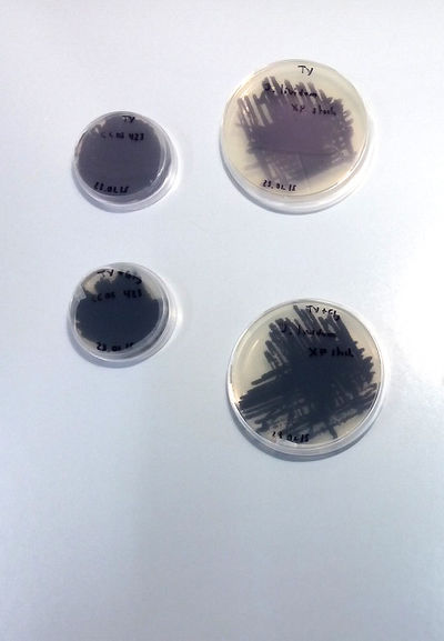
Nice! J.Lividum are growing nicely on a poor medium and scarily growing in poor TY + 1%Glycerol (86%). Although nicer color is showing with poor TY.
As in TY is growing fast enough and very nicely, we decided to continue the liquid medium with this configuration.
We put them 24h. on the shaker at 170rpm and 27°C.
27/01/2015
Après 24 heures a 27°C et 170 rpm on peut dire que on voit pas le violacein dans la bouteille. Donc on va essayer de ajouter Glycerol 1% et voir comment ils se comportent.
05/02/15
First Extraction following the article Pantanella-F_2007: [1] Abstract
Aims: To analyse the environmental stimuli modulating violacein and biofilm
production in Janthinobacterium lividum.
Methods and Results: Violacein and biofilm production by J. lividum
DSM1522T was assayed in different growth conditions. Our data suggest that
violacein and biofilm production is controlled by the carbon source, being
inhibited by glucose and enhanced by glycerol. J. lividum produced violacein
also in the presence of different sub-inhibitory concentrations of ampicillin. As
opposite, the production of N-acylhomoserine lactone(s), quorum sensing
regulators was shown to be positively regulated by glucose. Moreover, violaceinproducing
cultures of J. lividum showed higher CFU counts than violaceinnonproducing
ones.
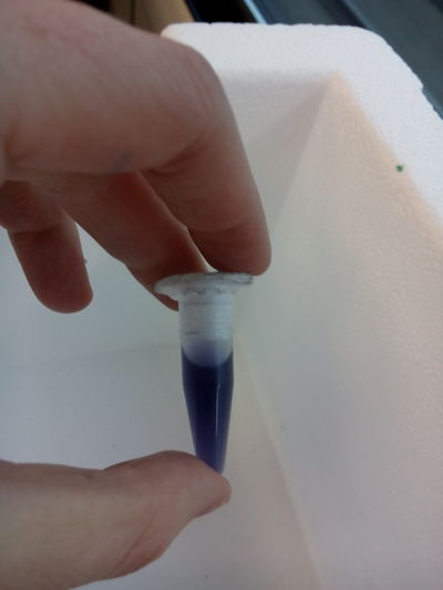
Extraction: Violacein extraction J. lividum was grown in 2 ml of LB or LB-U or LB-Y in 24-well plates. At regular time intervals, bacterial cells from a single well were harvested by centrifugation (16 000 g for 20 min), and treated for violacein extraction. Violacein extraction was carried out according to previously described methods (Blosser and Gray 2000; Matz et al. 2004). Briefly, bacterial cells were lysed with 10% sodium dodecyl sulfate. After 5 min at room temperature, water-saturated butanol was added (1 : 2 v/v) and vortexed. After centrifugation (16 000 g for 10 min) the upper phase, containing violacein, was collected. The extracted violacein was quantified using a spectrophotometer (OD585). The extinction coefficient of violacein (0Æ05601 ml lg)1 cm)1) was used for quantitative calculations (Mendes et al. 2001).
Note: We made the culture on TY and TY + Glycerol
SOME CONCLUSIONS:
TY and TY + Gly is perfect for J.Lividum (Violet)
MSA, TY not working for M.luteus (Yellow)
LB + 4gr. plus of "food" (yeast and peptone) neither. MLuteus grow better on LB agar and liquid medium at 37°C (maybe not well calibrated machine...)
10/02/15
Building the FilaBot DIY Kit, an extruder for plastics. 40 hours of intense work. Thank Michael for the moral support and electronic supervision.
12/02/15
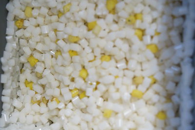
First attempts with Filabot extruder! ----> I've put the natural white pellets on the machine and verted the bio+ink. I have some problems with the porosity of the plastics, gonna try mabe ask an expert to know how to solve this.
The problem seems to be solved almost, if the pigments are added BEFORE putting them into the extruder let them dry (because of the extraction they already have alcohol so they dry fast) for a around 30min at room T°.
The result:
We could start printing with our Pr(ink)t as soon as I get to assembly and connect it!
Related Links
- Test Colonies Online FREE
- hackpad for the Prinkt plastic project
- Exhibition 2015 Milano fuori salone
- Bioink project of la paillasse
- Waag Society Workshop
Resources
